
A consolidated analytical review of four key works (2012, 2015, 2024, 2026)
Prepared from open scientific publications
Introduction. Why this topic is a revolution in neuroscience
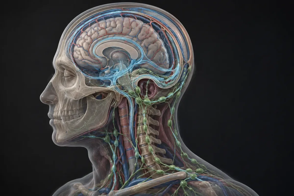
For nearly two hundred years, textbooks of anatomy and neurology maintained an ironclad assertion: the central nervous system (CNS) — the brain and spinal cord — does not possess a classical lymphatic system. Lymphatic vessels, which permeate virtually every other organ in the body and are responsible for returning interstitial fluid to the bloodstream, immune surveillance, and the removal of large molecules, supposedly terminate at the very base of the skull. Inside the brain, it was believed that cerebrospinal fluid (CSF) circulates in a closed circuit of the ventricles and subarachnoid space, while spent proteins are disposed of locally — by microglia and other resident cells. This model was elegant, internally consistent, and, as it turned out in the 2010s, fundamentally wrong. Over the past fourteen years — from 2012 to 2026 — four landmark papers were published, each of which rewrote the corresponding chapter of neuroanatomy. These works are not isolated: they form a unified chain in which each subsequent paper builds on the predecessors and fills the gaps they left behind. Understanding the fourth (Bhatt et al., 2026) without knowledge of the first three (Iliff 2012; Louveau and Aspelund 2015; Yoon 2024) is impossible — it would simply become a description of strange stained vessels on cervical preparations. Therefore, this text is structured as a single narrative: a historical exposition, four key papers, the unifying "Cerebrolymph" hypothesis, a critical analysis of limitations, and finally, clinical implications. This field is currently at a stage that in the philosophy of science is called a "paradigm shift" according to Thomas Kuhn: accumulated facts no longer fit the old theory, the new one has not yet stabilized, and each new preparation or mouse experiment can radically alter the picture. This makes the topic fascinating not only for specialists but for anyone interested in how scientific knowledge works — how dogma becomes a question, a question becomes a hypothesis, and a hypothesis becomes a map.
Brief roadmap
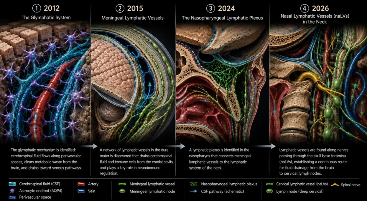
- 2012 — Maiken Nedergaard's laboratory in Rochester. Jeff Iliff publishes a paper opening up the glymphatic system: an internal "drainage" system of the brain, structured not like a conventional lymphatic network but as perivascular channels along blood vessels.
- 2015 — two independent groups (Louveau in Virginia and Aspelund in Helsinki) simultaneously demonstrate that the dura mater — the meninges — contains true classical lymphatic vessels with a full complement of endothelial markers. The dogma of CNS immune isolation collapses for good.
- 2024 — Yoon et al.'s work in Nature mapped the nasopharyngeal lymphatic plexus — a specific anatomical "exit" from the skull through which CSF and immune cells leave the cranial cavity and travel to the cervical lymph nodes.
- 2026 — Bhatt et al.'s paper in Cellular and Molecular Neurobiology describes lymphatic vessels in the cervical spine, directly at the boundary between the CNS and PNS. This is the missing anatomical segment between meningeal lymphatic vessels and deep cervical lymph nodes.
Historical background. Twenty-three centuries of misunderstanding
The idea that some "milky" or "whitish" fluid, distinct from blood, circulates in the body dates back to Hippocrates (460–375 BC), who described the so-called ichor — a transparent fluid flowing through the lumens of vessels. Aristotle (384–322 BC) went further and observed fibrous, fluid-containing structures between blood vessels and nerves. These early observations were qualitative, not anatomical in the modern sense — true lymphology only begins in the seventeenth century. The French physician Jean Pecquet (1622–1674) in 1651 identified the thoracic duct (ductus thoracicus) — the body's main lymphatic collector, through which lymph from the entire lower trunk, the left side of the head, and the left arm returns to the venous circulation at the so-called left venous angle, between the left subclavian and left internal jugular veins. Pecquet showed that lymph flows not to the liver (as ancient authors believed) but into the venous system. The Danish anatomist Niels Stensen (Nicolaus Steno, 1638–1686) described not only the thoracic duct and left jugular lymphatic trunk but also cervical lymphatic vessels — that is, the very tubular infrastructure of the neck through which we today trace the exit of fluid from the skull. The most prominent figure in the prehistory of this topic is the Italian anatomist Paolo Mascagni (1755–1815). In the late eighteenth century, he performed mercury injections into the lymphatic vessels of cadavers and obtained preparations of remarkable detail for the time. Among his findings were lymphatic vessels in the human dura mater — structures that we now call meningeal lymphatic vessels (MLVs). His anatomical wax models of dural lymphatic vessels are still preserved in the Josephinum medical museum in Vienna, Austria. The eighteenth century, therefore, already "knew" about cerebral lymphatic vessels.
"From Mascagni to Louveau — 228 years. Nearly a quarter of a millennium was needed to return to science what was already visible in wax models."
— From the commentary on Bhatt et al., 2026. But in the nineteenth and especially the twentieth century, this finding was "forgotten" under the pressure of skepticism. Large-scale anatomical mapping of the human lymphatic system almost completely ceased — anatomists switched to microscopic histology and new instrumental methods. In twentieth-century works (Lecco 1953; Földi et al. 1966; Lukić et al. 2003), descriptions of lymph-like structures in the dura mater and hints of connections between the CNS and peripheral lymphatic pathways periodically appeared, but these reports remained marginal: classical histology with conventional stains (hematoxylin–eosin) poorly distinguishes thin-walled lymphatic vessels from venules, and convincing immunohistochemical markers for lymphatic endothelium did not exist until the late 1990s. As a result, by the beginning of the twenty-first century, textbooks had solidified a model in which CSF circulates in a closed circuit of the ventricles and subarachnoid space, is reabsorbed into venous sinuses through arachnoid granulations (Pacchioni's granulations), and the brain as a whole is considered an "immune-privileged" organ — that is, protected from the immune system by the blood-brain barrier. This model explained much: why transplanted brain tumors are often not rejected; why the systemic immune response poorly penetrates the CNS; why the brain has no visible lymphatic vessels. But it did not explain where exactly the spent proteins go — especially β-amyloid, the accumulation of which underlies Alzheimer's disease. And this "waste question" became the starting point of the revolution in the 2010s.
Part I. Iliff et al., 2012 — discovery of the glymphatic system
Context of the work
In the early 2010s, Maiken Nedergaard's laboratory at the University of Rochester (New York State, USA) was conducting research on fluid movement in brain parenchyma. The young researcher Jeffrey J. Iliff was working with two-photon microscopy — a method that allows observation of fluorescent molecules in living tissue at depths of several hundred micrometers. The Nedergaard group posed a simple question: if the brain does not have lymphatic vessels, how then are large molecules dissolved in interstitial fluid removed — for example, β-amyloid (Aβ), a peptide of 40–42 amino acids whose aggregation into plaques leads to Alzheimer's disease? Experiments on mice with labeled Aβ showed that it disappears from the parenchyma remarkably quickly — within hours, not days. But exactly where it went remained unclear.
What exactly was discovered
In the paper Iliff JJ, Wang M, Liao Y, et al. (2012) "A paravascular pathway facilitates CSF flow through the brain parenchyma and the clearance of interstitial solutes, including amyloid β," published in Science Translational Medicine (volume 4, article 147ra111), a fundamentally new system was described. It was named the glymphatic system (from glia — glial cells, auxiliary cells of the CNS — and lymphatic). The essence of the discovery was as follows. Within the brain, there exist perivascular spaces — gaps between the wall of a blood vessel (arteries, arterioles, capillaries, veins) and the surrounding neural tissue. At cerebral arteries, these spaces are lined on the outside by astrocyte processes — the so-called astrocyte endfeet — which cover the vessel almost entirely, leaving only narrow gaps. Through these perivascular channels, CSF entering from the subarachnoid space penetrates deep into the parenchyma, bathes the neurons and interstitium, exchanges with interstitial fluid, and then returns to the subarachnoid space via venous perivascular channels. This circulation, unlike classical lymphatic drainage, does not use closed lymphatic vessels with valves — it is driven by arterial pulsations, pressure gradients, and, as was later discovered, respiratory rhythms. Methodologically, the key breakthrough was the use of two-photon fluorescence microscopy in live mice: through a cranial window, researchers observed in real time the movement of labeled CSF (with a fluorescent dye) and labeled β-amyloid. The dye entered the parenchyma along arteries, spread through the interstitium, and then exited along veins. β-amyloid was cleared by the same route, and its clearance rate depended on the presence of aquaporin-4 (AQP4) — a water channel protein localized specifically in astrocyte endfeet. In mice with genetic Aqp4 knockout, β-amyloid clearance was reduced by approximately 55–65%. This was direct functional evidence: the glymphatic system works, and it works through perivascular glial channels.
Sleep and brain cleansing
A year after the first publication, in Science (Xie et al., 2013, volume 342, pp. 373–377), the same laboratory demonstrated a second effect that instantly made the topic famous: glymphatic clearance is active during sleep. In sleeping mice, the interstitial space in the brain expands by approximately 60% (due to a reduction in the volume of the cells themselves), and β-amyloid clearance accelerates twofold compared to wakefulness. This explained why chronic sleep deprivation correlates with an increased risk of neurodegenerative diseases — the brain literally "cannot keep up" with washing out its own metabolic waste.
"We sleep so the brain can wash itself. The glymphatic system works mainly at night — this was the slogan that swept through all popular science publications in 2013."
— Popular account of Xie et al., 2013
What the glymphatic system did NOT explain
The 2012 discovery solved half the problem — it showed how fluid moves within the brain. But the second, equally important question remained: exactly where does it exit the skull? The glymphatic system is internal; it redistributes CSF between the parenchyma and the subarachnoid space. But it did not describe the final "exit" into the peripheral lymphatic system. Meningeal lymphatic vessels were still considered nonexistent. Therefore, the logical next step was to search for the peripheral link — and this came in 2015.
Full bibliographic reference
Iliff JJ, Wang M, Liao Y, Plogg BA, Peng W, Gundersen GA, Benveniste H, Vates GE, Deane R, Goldman SA, Nagelhus EA, Nedergaard M. (2012) A paravascular pathway facilitates CSF flow through the brain parenchyma and the clearance of interstitial solutes, including amyloid β. Science Translational Medicine, 4(147):147ra111. doi:10.1126/scitranslmed.3003748.
Part II. Louveau and Aspelund, 2015 — meningeal lymphatic vessels
Two independent groups, one discovery
In 2015, in the journals Nature and the Journal of Experimental Medicine, practically simultaneously — with a gap of only a few weeks — two papers appeared, both demonstrating the same thing: the mouse dura mater contains classical lymphatic vessels. This is a rare occurrence in science, when two independent groups, unaware of each other's work, arrive at the same result using different methods. Antoine Louveau's work at Jonathan Kipnis's laboratory at the University of Virginia appeared in Nature (volume 523, pp. 337–341). Aleksi Aspelund's work from the Institute of Biomedicine, University of Helsinki, appeared in the Journal of Experimental Medicine (volume 212, pp. 991–999).
What exactly was found
In the dura mater — the outermost of the three meninges, lining the inner surface of the skull — along the superior sagittal sinus and other dural sinuses (large venous collectors), there are true lymphatic vessels. They possess a full complement of lymphatic endothelial markers: PROX1 (a transcription factor specifying the lymphatic fate of a cell), LYVE-1 (a hyaluronan receptor), VEGFR-3 (the lymphatic growth factor VEGF-C receptor), podoplanin (a transmembrane protein recognized by the D2-40 antibody), and CCL21 (a chemokine that attracts dendritic cells and T lymphocytes). These vessels have valves, through which fluid moves unidirectionally — from meningeal spaces toward the skull exits, to the deep cervical lymph nodes (dcLNs).
Why was this not seen before
The reason is technological. Lymphatic vessels in the dura mater are very thin-walled, collapsed in preparations, and difficult to distinguish by conventional histology. To visualize them, specific antibodies against lymphatic markers (PROX1, LYVE-1, VEGFR-3, D2-40) are needed, which only became widely available in the 2000s. Moreover, the "dogma" about their absence was so deeply entrenched in textbooks that nobody simply looked. Legend has it that the Kipnis laboratory stumbled upon these vessels accidentally — while studying T lymphocyte trafficking in the mouse meninges.
Functional data from Aspelund
Aspelund's group went beyond mere description. They used mice with genetic knockout of the transcription factor PROX1 in endothelium (which prevents lymphatic vessel development) and obtained animals lacking meningeal lymphatic vessels (MLVs). In such mice: (1) CSF clearance from the subarachnoid space to deep cervical lymph nodes was reduced by 50–70%; (2) β-amyloid accumulation in brain parenchyma was accelerated; (3) in aged mice, MLVs "deflate" — their lumen narrows, density decreases, and clearance worsens further, correlating with cognitive decline in memory tests; (4) administration of exogenous VEGF-C — a lymphatic growth factor — to old mice restored MLVs and improved amyloid clearance. This was a direct demonstration: MLVs are a functionally significant waste clearance pathway from the brain, and they can be therapeutically modulated.
The immunological perspective from Louveau
In parallel, Louveau's group added an immunological perspective. In mice with genetically defective MLVs, T lymphocytes infiltrated the brain less effectively, and in experimental models of multiple sclerosis (an autoimmune CNS disease, the EAE model — experimental autoimmune encephalomyelitis), symptoms were milder. This overturned the very concept of brain immune privilege: the brain is not absolutely sealed off from the immune system; it has a very delicate, controlled interface, and MLVs are the key link in this interface. Through MLVs, immune cells can enter the brain (which is important in autoimmune diseases), and through them the brain also "reports" to the immune system about its condition.
What remained unclear
The 2015 papers left one major gap. Meningeal lymphatic vessels were found inside the skull. They drain fluid into deep cervical lymph nodes — this was shown. But exactly how does fluid exit the skull to reach the cervical lymph nodes? Where are the specific anatomical conduits connecting the cranial circuit with the peripheral lymphatic system? For ten years this was debated at conferences — and only in 2024 did a map of these conduits appear. But more on that in the next section.
Full bibliographic references
Louveau A, Smirnov I, Keyes TJ, Eccles JD, Rouhani SJ, Peske JD, Derecki NC, Castle D, Mandell JW, Lee KS, Harris TH, Kipnis J. (2015) Structural and functional features of central nervous system lymphatic vessels. Nature, 523(7560):337–341. doi:10.1038/nature14432. Aspelund A, Antila S, Proulx ST, Karlsen TV, Karaman S, Detmar M, Wiig H, Alitalo K. (2015) A dural lymphatic vascular system that drains brain interstitial fluid and macromolecules. Journal of Experimental Medicine, 212(7):991–999. doi:10.1084/jem.20142290.
Part III. Yoon et al., 2024 — the nasopharyngeal lymphatic plexus
Context of the work
After 2015, the two ends of the pathway became clear: intracranial meningeal lymphatic vessels and cervical lymph nodes. But the middle — the specific anatomical transition between them — remained terra incognita. It was known that in mice CSF exits the skull somehow through the skull base, but the precise "portal" remained undescribed. In January 2024, a paper in Nature (volume 625, pp. 768–777) closed this gap in the mouse and provided the first confirmations in humans.
The main discovery
Yoon J-H et al. (2024) "Anatomical and functional analyses of the meningeal lymphatic vasculature" described a specific anatomical hub through which CSF exits the skull. This is the nasopharyngeal lymphatic plexus (NLP) — a network of lymphatic vessels in the soft tissues of the nasopharynx, located directly beneath the skull base. Thus we have a pathway: subarachnoid space → meningeal lymphatic vessels → exit through the skull base (along the olfactory, trigeminal, and other cranial nerves) → nasopharyngeal plexus → deep cervical lymph nodes. Yoon and colleagues showed that this is not an abstract pathway but a highly specific one: they visualized it in mice using fluorescent tracers and confirmed it in humans with MR lymphangiography.
Methods and results
The researchers used several complementary approaches. First, genetically labeled mice (Prox1-GFP) with fluorescent lymphatic endothelial cells, enabling detailed mapping of the lymphatic architecture at the skull base. Second, injections of fluorescent tracers into the cisterna magna (the large cistern of the subarachnoid space) with subsequent observation of their exit from the skull. Third, selective surgical ablation of the nasopharyngeal plexus with assessment of disrupted CSF drainage. Fourth, MR lymphangiography in humans with contrast agent injected into the soft tissues of the neck to visualize the lymphatic network of the skull base. The main quantitative findings: the nasopharyngeal plexus accounts for approximately half of CSF drainage from the skull in mice. After its surgical removal, CSF appeared in cervical lymph nodes 40–50% more slowly, and intracranial pressure rose slightly. Age-related involution of the plexus correlated with impaired β-amyloid clearance. In older human subjects, MRI showed reduced size and density of the nasopharyngeal plexus, consistent with the well-established increased risk of neurodegeneration in the elderly.
Clinical significance
Yoon's work provided a concrete anatomical target for therapeutic interventions. If the nasopharyngeal plexus is functionally important for CSF drainage, its stimulation (for example, with the same VEGF-C) or its preservation during age-related involution could slow the development of Alzheimer's disease and other neurodegenerative conditions. The first clinical speculations emerged regarding a connection between chronic rhinitis, nasopharyngeal allergic diseases, and neurodegeneration — but these are still hypotheses requiring direct testing.
What remained unclear
Yoon and colleagues resolved the question of the "skull exit." But what happens next, in the neck? Which specific lymphatic vessels carry fluid from the skull base downward through the neck to the deep cervical lymph nodes? Do lymphatic vessels exist along the cervical spinal nerves — that is, at the boundary between the CNS and the peripheral nervous system (PNS)? This anatomical territory remained unexplored. And here, in 2026, Bhatt et al.'s work appears as the final piece of the puzzle.
Full bibliographic reference
Yoon J-H, Nguyen KP, Nhan HTT, et al. (2024) Anatomical and functional analyses of the meningeal lymphatic vasculature. Nature, 625(7996):768–777. doi:10.1038/s41586-023-06799-9.
Part IV. Bhatt et al., 2026 — lymphatic vessels of the cervical spine
Bibliographic summary
Bhatt SS, Coker CR, Bagian LK, Tornatore CS, Kulkarni JA, Um JH, Weatherdon ET, Hallowell CR, Akhtarzandi-Das AM, Dreger-Tretheway MN, Casely AJ, Deale EV, DeLeonibus SF, Li H, Hakeem JS, Bhatt SV, Talwar D, Olabosipo JO, Caputo JM, Harris BT. (2026) Postmortem Evidence of Lymphatic Vessels Located at the Boundary of Central and Peripheral Nervous Systems in the Cervical Spine. Cellular and Molecular Neurobiology. doi:10.1007/s10571-026-01744-4. Published 5 June 2026. Corresponding author: Brent T. Harris, Georgetown University, Washington, District of Columbia.
Purpose and logic of the study
The logic of the work is as follows. From the 2012–2024 studies, we knew about the intracranial link (MLVs) and the nasopharyngeal exit (NLP). But the neck — the segment between the skull base and the clavicle — remained anatomical "terra incognita" with respect to lymphatic vessels associated with spinal nerves. The authors posed the question: do lymphatic vessels exist that accompany cervical spinal nerves as they exit the vertebral canal through the intervertebral foramina? In other words, is there lymphatic infrastructure at the boundary between the CNS (the spinal cord and its meninges within the vertebral canal) and the PNS (spinal nerves and ganglia outside the canal)? This zone is called the craniovertebral junction and the cervical intervertebral foramina.
Methodology. Part 1: retrograde dye injection
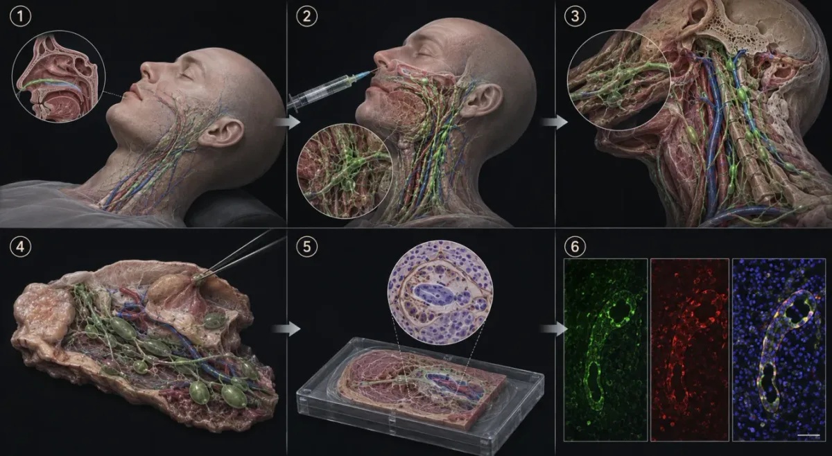
The study was conducted on nine cadaveric donors from the Georgetown University anatomical donor program. All donors had been fixed in formalin — this is standard practice for anatomical studies, but it creates a serious technical problem: formalin fixes tissues, thickens vessel walls, glues valves shut, and makes it practically impossible to inject anything into a lymphatic vessel in this state. The authors found an elegant workaround. First, the thoracic duct (ductus thoracicus) — the body's main lymphatic collector — was identified in the thoracic cavity. It was traced from the abdominal cavity to the point of insertion at the left venous angle — the site where the left internal jugular vein joins the left subclavian vein. To prevent retrograde flow of dye into the venous system, the left brachiocephalic, subclavian, and internal jugular veins were clamped with hemostatic forceps distal to the duct's insertion point. The duct was then transected at two points. At point 1 (at the level of T5), 20 mL of 5% hydrogen peroxide was injected — the "primer." Hydrogen peroxide works as a chemical dilator: oxygen bubbles gently expand the vessel lumen, dissolve small thrombi and clots, and clean the walls. A 30-gauge needle, ½ inch length, manual low-pressure injection, without visible excessive distention or rupture. At point 2 (at the left jugular lymphatic trunk, distal to its junction with the thoracic duct), a repeat dose of peroxide was administered, followed by the main injection — a 3:1 mixture of 5% H₂O2 and green Rit dye (total 50 mL).
"A wooden block, nightfall, and gravity. No high technology — just patience and physics."
— From the protocol description in Bhatt et al., 2026. Why green? The authors honestly describe a process of trial and error. Blue dye proved unsuccessful — it was difficult to distinguish from venous vessels, which are also bluish. Black dye in mixture with peroxide produced a precipitate. Green was optimal: brightly visible, not confused with veins, and did not precipitate. If during injection a lymphatic vessel ruptured and dye leaked beyond the lumen, the leak was sealed with ordinary superglue (cyanoacrylate), allowed to dry, and the procedure continued. If superglue occluded the lumen, the vessel was transected more proximally and the procedure continued at a new segment. After injection, the body was left overnight in the following position: a wooden block under the upper chest, the neck maximally extended backward (hyperextension), the head tilted back. Gravity over 12–16 hours slowly pushed the dye cranially — upward, toward the skull base. The authors honestly acknowledge that retrograde filling is inherently incomplete: formalin thickens walls and valves, and the neck contains many valvular stations that impede reverse flow. Therefore the method only illuminates those lymphatic pathways that remained patent. This is not a complete lymphangiogram but an opportunistic indicator.
Methodology. Part 2: sample collection and immunohistochemistry
The next day, the body was dissected. Two main visualization approaches were used. First, the calvarium was removed to view the skull base from above. Second, a cervical tissue flap was created preserving the stained vessels, the neck was transected, and the head was bisected along the midline to observe dye distribution in midline structures. Any stained vessels that clearly emerged from the cranial or spinal cavities were excised at the most proximal entry point along with surrounding tissue for histological analysis. Collected samples were fixed in 70% ethanol; bony portions were decalcified with formic acid. Standard hematoxylin and eosin (H&E) staining was used for tissue morphology. For confirmation of lymphatic identity, immunohistochemistry with the D2-40 antibody — a monoclonal antibody against podoplanin, a transmembrane protein of lymphatic endothelium — was employed. Paraffin blocks were cut into 4 μm sections and mounted on charged slides. Deparaffinization, rehydration, and epitope retrieval were performed on a Dako PT Link instrument with EnVision FLEX Target Retrieval Solution High pH (9.0) at 97 °C for 20 minutes. Immunostaining was performed on a Dako Autostainer Link 48 with a ready-to-use mouse monoclonal anti-podoplanin antibody (D2-40, clone D2-40, Dako IR072). Incubation: peroxidase blocking 5 minutes, D2-40 20 minutes, FLEX Mouse Linker 20 minutes, FLEX HRP 20 minutes, FLEX DAB Substrate-Chromogen 10 minutes, FLEX Hematoxylin 5 minutes. Between steps — washing with EnVision FLEX Wash Buffer. Expected staining pattern: membranous, in lymphatic endothelium.
Results. What was found in donors
Of nine donors, stained vessels were found in four (Donor 1, 2, 3, 4) in the vertebral canal or at the cervical intervertebral foramina, which were collected for histological analysis. In the first three donors, staining was confirmed by immunohistochemistry (D2-40-positive); in the fourth, it was only macroscopic without IHC (and therefore considered preliminary).
Donor 1 (black dye)
Two positive localizations. The first — in the retrodental space, posterior to the odontoid process (dens axis, the second cervical vertebra) and anterior to the dura mater at the craniovertebral junction. This is the uppermost point of the cervical spine — where the skull transitions to the vertebral column. The second — at the level of C2–C3, where a stained vessel followed the C3 spinal nerve as it passed through the intervertebral foramen. Both samples were D2-40-positive.
Donor 2 (green dye)
One positive sample from the lateral region of the neck, in the interscalene triangle, near the brachial plexus. A stained vessel accompanied a spinal nerve heading toward an intervertebral foramen. D2-40-positive.
Donor 3 (green dye)
The richest donor — four positive samples. Two distinct stained perineural samples from adjacent cervical roots in the lateral neck (presumably components of the brachial plexus) — both D2-40-positive. Two additional samples from the retropharyngeal region (posterior to the pharynx), also D2-40-positive. All four samples show thin-walled luminal structures with membranous podoplanin staining, morphologically corresponding to lymphatic vessels.
Donor 4 (green dye)
Macroscopically positive dye uptake bilaterally — on left and right cuts. Stained areas: nasopharyngeal region below the sphenoid sinus and right buccal mucosa. These samples were not processed for histology or immunohistochemistry and are therefore interpreted as preliminary.
Summary table by donors
• Donor 1: black dye; vessel at CNS/PNS boundary — positive; 2 samples collected; D2-40+ — 2. • Donor 2: green dye; vessel at CNS/PNS boundary — positive; 1 sample collected; D2-40+ — 1. • Donor 3: green dye; vessel at CNS/PNS boundary — positive; 4 samples collected; D2-40+ — 4. • Donor 4: green dye; vessel at CNS/PNS boundary — positive; samples collected: n/a; D2-40+ — n/a. In total, in 4 of 9 donors (44%), it was possible to retrogradely fill and confirm D2-40-positive vessels along cervical spinal nerves at the level of the intervertebral foramina. The authors introduce the designation naLVs for these vessels — nerve-adjacent lymphatic vessels.
Anatomical localizations of naLVs
In summary across all donors, D2-40-positive lymphatic vessels were found in the following anatomical compartments: - Paradural lymphatic vessels in tissues directly ventral to the dura mater at the craniovertebral junction — that is, at the very apex of the cervical spine, where the first cervical vertebra (atlas) articulates with the skull. - Lymphatic vessels adjacent to the perineurium in bundles exiting through intervertebral foramina (at levels C2–C3, and more caudally in the brachial plexus region). - More superficial lymphatic vessels in the surrounding soft tissues of the neck — the retropharyngeal region, lateral cervical region. - Macroscopically stained (but not IHC-confirmed) areas at the skull base — nasopharynx below the sphenoid sinus, buccal mucosa.
Authors' hypothesis. What these vessels may mean
The main anatomical conclusion of the work: lymphatic vessels accompany cervical spinal nerves as they exit the vertebral canal and pass through the intervertebral foramina. They are D2-40-positive, thin-walled, with membranous podoplanin staining — morphologically corresponding to lymphatic vessels. Since dye was injected retrogradely (from the thoracic duct and jugular trunk), rather than anterogradely (from the subarachnoid space), only those lymphatic channels that are anatomically continuous with the peripheral lymphatic network were filled. This means that the stained cervical dural and epineural vessels are part of the functioning peripheral lymphatic network, not isolated, blindly terminating channels. The authors emphasize an important limitation: D2-40 also marks the perineurium (the peripheral nerve sheath), and some D2-40-positive profiles are located immediately adjacent to the perineural sheaths. Therefore, they are interpreted as intraepineural lymphatic vessels tightly apposed to the perineurium, but they cannot fully exclude that some of the staining belongs to the perineurium itself. For a definitive resolution, additional markers (LYVE-1, PROX1, SMA) are needed. Also critically important: the study did NOT directly visualize CSF or intradural lymphatic channels. Any conclusions regarding CSF drainage or glymphatic clearance should be regarded as hypotheses requiring dedicated functional and imaging studies. The authors do not show that fluid actually flows through these vessels from the brain — they show that the vessels are present, that they are lymphatic by markers, and that they are anatomically continuous with the peripheral lymphatic network.
Part V. The "Cerebrolymph" hypothesis. A unified scheme of neurofluid homeostasis
The most ambitious theoretical construct of Bhatt et al., 2026 is the authors' proposed "Cerebrolymph" hypothesis. This is an attempt to assemble all the puzzle pieces — Iliff's, Louveau-Aspelund's, Yoon's, and the authors' own findings — into a unified scheme. The full cycle of neurofluid homeostasis appears, according to the authors, as follows.
Six stages of the complete cycle
• Stage 1. CSF production. Oxygen-rich blood enters the cranial cavity through the internal carotid and vertebral arteries, perfuses the choroid plexus of the ventricles via choroidal branches, and is there filtered into CSF. At this stage, the blood-CSF barrier (BCSFB) operates. • Stage 2. Circulation through the ventricles. CSF circulates through the ventricular system (lateral → third → fourth). Circumventricular organs (CVOs) — specialized structures lacking a blood-brain barrier — line the ventricular walls and regulate CSF composition through sensory and secretory functions. Between CVOs and the ventricular CSF lies a layer of tanycytes — specialized ependymal cells with tight junctions forming an additional barrier. • Stage 3. Entry into the subarachnoid space. Through the median aperture (foramen of Magendie) and lateral apertures (foramina of Luschka) of the fourth ventricle, CSF enters the subarachnoid space. • Stage 4. Glymphatic exchange. In the subarachnoid space, CSF participates in glymphatic exchange: arterial perivascular influx, driven by cardiac pulsations and vasomotion, delivers CSF into the parenchyma through astrocyte endfeet at the glia interface, facilitating the clearance of interstitial dissolved substances. Venous perivascular efflux, modulated by respiratory rhythms, returns substance-rich fluid to the subarachnoid space. • Stage 5. Drainage via two parallel pathways. At the boundary of the subarachnoid space and the dura mater, cleaned fluid exits through two coordinated pathways: (1) reabsorption into dural sinuses via arachnoid granulations, and (2) uptake by meningeal lymphatic vessels via arachnoid granulations and arachnoid cuff exit points (ACE). The authors introduce the term "cerebrolymphatic barrier" (CLB) to denote the fraction of CSF that drains into MLVs and the lymphatic fraction that can dynamically interact with the subarachnoid pool. • Stage 6. Extracranial continuation. The present work (Bhatt et al., 2026) considers naLVs — lymphatic vessels located adjacent to cervical nerves — as the extracranial continuation of meningeal lymphatic fluid drainage, draining into the deep cervical lymph nodes (dcLNs). This stage is supported by D2-40 immunohistochemical data presented in the paper.
Bidirectionality of the cerebrolymphatic barrier
An interesting nuance of the hypothesis: ACE points (arachnoid cuff exit points) — bidirectional interfaces along bridging veins, where breaks in the arachnoid barrier allow both drainage of CSF into the dura mater and selective molecular or cellular migration from the dura mater into the subarachnoid space. This links CNS clearance and immune communication into a single bidirectional interface. Direct evidence of retrograde meningeal lymphatic flow is emerging but still limited: in an in vivo mouse study (Ramos-Zaldívar et al., 2024), nanoparticles were injected into deep cervical lymph nodes and their movement was tracked back to the meninges and brain, explicitly identified as "retrograde directed flow." Human MRI studies (Kuo et al., 2018) showed that flow in dorsal MLVs adjacent to the superior sagittal sinus is directed posterior-to-anterior — opposite to and against the direction of venous blood flow.
What the hypothesis does NOT claim
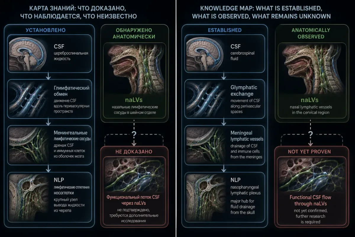
The authors very carefully emphasize: "Cerebrolymph" is a conceptual anatomical framework, not a proven drainage pathway. Each individual component of this cycle is supported by established literature, but their assembly into a unified scheme is precisely a hypothesis requiring functional verification. The work of Bhatt et al., 2026 demonstrates the anatomical existence of naLVs but does not show actual fluid flow through them.
Limitations of the Bhatt et al., 2026 study
Technical limitations
The first and foremost limitation is the use of formalin-fixed, embalmed donors with retrograde dye injection. This yields only an incomplete and nonphysiological picture of patent channels; the authors could not assess dynamic flow, valve function, or in vivo filling patterns. Of the nine donors, staining was successful in only four — in the rest, either the dye failed to ascend, the vessels ruptured, or leakage occurred beyond the lumen. This means the method illuminates only a portion of the real lymphatic network, not the whole of it. Second: the authors did not directly trace continuity between the subarachnoid CSF spaces and the described D2-40-positive vessels. Therefore, observations should be interpreted as an anatomical description of lymphatic-like vessels at cervical CNS exit sites, rather than as functional proof of CSF drainage. Future studies on fresh or lightly fixed tissue, with systematic H&E correlation and multiple lymphatic and neuronal markers (LYVE-1, PROX1, SMA), will be needed for definitive identification of vessels at these boundary sites.
Resource limitations
Third: resource limitations. Limited dissection time, a small number of cadaveric donors (nine is few for an anatomical study), and limited funding for comprehensive histological and immunohistochemical processing all prevented analysis of all potentially relevant samples. The novelty and evolving nature of the injection protocol meant that some preparations were less informative, as the technique was optimized by trial and error. Anatomical variability among donors further limits the generalizability of results. Fourth: access to lymphatic vessels through dissection frequently required removal of surrounding tissue, which could disrupt their natural relationships or damage delicate vessels. Dye migration failures could also reflect unnoticed intraluminal obstructions or suboptimal primer/dye volumes. Since samples were fixed in formalin and processed after dye injection, some intraluminal dye was likely lost or redistributed during fixation, decalcification, and sectioning.
The D2-40 specificity problem
Fifth and very important methodological note: D2-40 (anti-podoplanin antibody) is not an absolutely specific marker for lymphatic endothelium. It also marks the perineurium — the connective tissue sheath around nerve bundles. Therefore, some D2-40-positive profiles in sections, particularly those immediately adjacent to the perineurium, may actually represent the perineurium itself rather than distinct lymphatic vessels. The authors acknowledge this and interpret their findings as "intraepineural lymphatic vessels tightly apposed to the perineurium," but cannot fully exclude that some staining belongs to the perineurium without additional markers (LYVE-1, PROX1, SMA). Sixth: although all D2-40-positive structures were morphologically assessed on standard sections, the paper does not systematically present paired H&E images for each immunostained vessel. This means that wall architecture and luminal profiles are consistent with lymphatic identity, but for definitive exclusion of periarterial or other non-lymphatic structures, additional markers and comprehensive H&E+IHC panels are needed in future work. Seventh: macroscopically stained skull bases and nasopharyngeal areas in Donor 4 were not subjected to IHC, so any conclusions about intracranial drainage from these sites should be considered preliminary.
Clinical and scientific implications
Alzheimer's disease and neurodegeneration
The most obvious clinical implication: if lymphatic drainage from the brain is functionally significant for clearing β-amyloid and tau protein (two key pathological proteins in Alzheimer's disease), then its stimulation could slow the development of neurodegeneration. Data already exist that in aged mice MLVs "deflate" and are less effective at drainage, correlating with cognitive decline. Administration of exogenous VEGF-C (a lymphatic growth factor) restores MLVs and improves amyloid clearance. The logical next step is clinical trials of VEGF-C or its analogs in Alzheimer's disease, but such trials do not yet exist. Alternative approaches include physical stimulation of lymphatic flow through breathing practices, posture, and physical activity.
Multiple sclerosis and autoimmune CNS diseases
A parallel story — multiple sclerosis and other autoimmune CNS diseases. If MLVs are the "entry door" for auto-reactive T lymphocytes into the brain, then their blockade could be therapeutically beneficial. In mice with genetically defective MLVs, symptoms of experimental autoimmune encephalomyelitis (EAE, a mouse model of multiple sclerosis) were milder. But translating this to the clinic is a complex task: systemic blockade of lymphangiogenesis has numerous side effects, and selective blockade of MLVs requires the development of tissue-specific delivery vehicles.
Cervical lymphatic vessels and neck pathologies
The discovery of naLVs (nerve-adjacent lymphatic vessels) by Bhatt et al. adds a new perspective to this. If lymphatic vessels along cervical spinal nerves are functionally significant for fluid drainage from the skull base, then any neck pathologies affecting these vessels could have remote neurological consequences. These could include: cervical osteochondrosis with nerve root compression; inflammatory diseases of the neck soft tissues; consequences of neck surgery (for example, lymph node dissection in oncology); prolonged flexed neck posture (so-called text neck from smartphone use). All these conditions could theoretically impair lymphatic flow through naLVs and, consequently, CSF drainage from the skull. Direct evidence is lacking, but it is anatomically plausible.
Side sleeping and posture
Several speculative but interesting implications. Side sleeping: in mice, glymphatic clearance is most efficient in the lateral position, less so on the back, and least on the stomach. This is a correlational study but an interesting hint for humans. Neck posture: prolonged flexed neck posture, as with smartphone use, could theoretically impair lymphatic flow through cervical naLVs. Direct evidence is lacking, but it is anatomically plausible. Breathing practices: venous perivascular efflux is modulated by breathing, and deep diaphragmatic breathing may improve CSF dynamics — this is indirectly supported by MRI studies.
Age-related involution
With age, MLVs "deflate" and become less effective at drainage. This is one of the possible mechanisms linking aging with neurodegeneration. If cervical naLVs also involute with age, this creates a double "drainage deficit": both in the skull and in the neck. Clinically, this could manifest as an increased risk of neurodegeneration in the elderly, which is consistent with epidemiology. But direct studies on large samples are needed — Bhatt et al.'s work describes only nine donors, which is too few to assess age-related dynamics.
Open questions and directions for future research
Anatomical studies
The first direction is expansion of the anatomical map. Bhatt et al.'s work covered only the cervical spine. But spinal nerves exit the vertebral canal at all levels — thoracic, lumbar, sacral. Do analogous naLVs exist along these nerves? If so, what is their density and their relationship with the peripheral lymphatic system? This is work for the next ten years, requiring large sample sizes of preparations and standardized protocols. Hundreds of preparations will be needed to confirm that naLVs are a normal component of human anatomy rather than a chance finding in four of nine donors. It will also be necessary to integrate high-resolution imaging with systematic H&E and multiplex immunohistochemistry for more precise identification of vessel identity and their smooth muscle coverage.
In vivo functional studies
The second direction is in vivo functional imaging in humans. Currently we have only postmortem anatomical data. Dynamic studies with labeled tracers and MR lymphangiography are needed to demonstrate that fluid actually flows through naLVs in living organisms. This is technically challenging: MR lymphangiography requires injection of contrast into the lymphatic system, which is invasive. An alternative is the use of endogenous contrast agents (for example, the FLAIR signal from CSF) or new imaging methods (low-field MRI, optoacoustic imaging).
Clinical trials
The third direction is clinical trials. Can lymphatic drainage be stimulated in Alzheimer's disease? Can it be blocked in multiple sclerosis? These are questions whose answers will not appear for at least 5–10 years. Early clinical data already exist suggesting that exercises improving venous return from the head (inversion therapy, certain forms of yoga) may improve cognitive function in the elderly — but these are not randomized controlled trials, and the link with lymphatic drainage is still hypothetical.
Comparative anatomy and evolution
The fourth direction is comparative anatomy. Do MLVs and naLVs exist in other mammals? In primates? In rodents? If so, how did they evolve? This is important for understanding how relevant mouse models (on which most of the work was done) are to humans. Data on differences already exist: in mice, MLVs are less developed than in humans, and their nasopharyngeal plexus is structured differently. This means that translating findings from mouse to human requires caution.
The glymphatic system and drug pharmacokinetics
The fifth direction, less obvious but practically important: understanding the glymphatic system may alter the pharmacokinetics of drugs acting on the CNS. If CSF moves through perivascular channels, then the residence time of a drug in the brain depends on glymphatic speed. And that speed depends on sleep, posture, age, and physical activity. This could explain why the same drug works differently in different patients. Future research may lead to "glymphatic-optimized" drug dosing regimens.
Key facts and figures for quick orientation
Timeline
1. 2012 — Iliff et al. — discovery of the glymphatic system. Sci Transl Med 4:147ra111. 2. 2013 — Xie et al. — glymphatic clearance is active during sleep. Science 342:373. 3. 2015 (May) — Louveau et al. (Nature 523:337) and Aspelund et al. (J Exp Med 212:991) — two independent papers on meningeal lymphatic vessels. 4. 2024 (January) — Yoon et al. — nasopharyngeal lymphatic plexus. Nature 625:768. 5. 2026 (June) — Bhatt et al. — lymphatic vessels of the cervical spine. Cell Mol Neurobiol. doi:10.1007/s10571-026-01744-4.
Key terms
• Glymphatic system — perivascular channels formed by astrocyte endfeet along brain arteries and veins. Through them CSF enters the parenchyma and exchanges with the interstitium, washing out spent proteins including β-amyloid. Active predominantly during sleep. • Meningeal lymphatic vessels (MLVs) — classical lymphatic vessels located in the dura mater along the sinuses. Possess a full complement of lymphatic endothelial markers: PROX1, LYVE-1, VEGFR-3, podoplanin (D2-40). First described by Mascagni (1787), confirmed by modern methods in 2015. • Nasopharyngeal lymphatic plexus (NLP) — a network of lymphatic vessels in the soft tissues of the nasopharynx, directly beneath the skull base. The main "exit" of CSF from the skull in mice. Described by Yoon et al., 2024. • naLVs (nerve-adjacent lymphatic vessels / nasal lymphatic vessels) — lymphatic vessels located adjacent to nerves, in the epineural and peridural compartments of the cervical spine. Described by Bhatt et al., 2026. • D2-40 — a monoclonal antibody against podoplanin, a transmembrane protein of lymphatic endothelium. Used for immunohistochemical identification of lymphatic vessels. Also marks the perineurium. • dcLNs — deep cervical lymph nodes, the terminal destination of lymphatic drainage from the meninges. • "Cerebrolymph" hypothesis — a unified anatomical conceptual framework describing the complete cycle of neurofluid homeostasis: CSF production → circulation through the ventricles → entry into the subarachnoid space → glymphatic exchange → drainage via meningeal lymphatic vessels → cervical naLVs → dcLNs. Proposed by Bhatt et al., 2026.
Figures from the Bhatt et al., 2026 study
- 9 — total number of cadaveric donors.
- 4 — number of donors with confirmed D2-40-positive vessels (44%).
- 5% — H₂O₂ concentration in the primer.
- 20 mL — volume of primer (H₂O₂) injection into the thoracic duct.
- 50 mL — total volume of dye mixture injection (3:1 H₂O₂ and green Rit).
- 12–16 hours — duration of overnight incubation with elevated chest.
- 4 μm — thickness of paraffin sections for IHC.
- 97 °C, 20 minutes — epitope retrieval conditions (HIER) for D2-40.
Figures from previous studies
- 55–65% — reduction in β-amyloid clearance in AQP4 knockout mice (Iliff 2012).
- 2× — acceleration of β-amyloid clearance during sleep compared to wakefulness (Xie 2013).
- 60% — expansion of brain interstitial space during sleep (Xie 2013).
- 50–70% — reduction in CSF drainage to dcLNs in mice lacking MLVs (Aspelund 2015).
- ~50% — contribution of the nasopharyngeal plexus to total CSF drainage from the skull in mice (Yoon 2024).
- 228 years — interval from Mascagni's description of MLVs (1787) to their modern confirmation (2015).
What is NOT shown in the Bhatt et al., 2026 study
- It is not shown that fluid actually flows through naLVs in living organisms.
- Continuity between the subarachnoid space and naLVs is not demonstrated.
- It is not shown that naLVs drain CSF specifically (as opposed to some other fluid).
- It cannot be excluded that some D2-40-positive staining belongs to the perineurium rather than to lymphatic vessels.
- Age-related dynamics of naLVs are not assessed (the sample of 9 donors is too small).
- Pathological states (neurodegeneration, multiple sclerosis, cervical pathologies) are not assessed.
Conclusion. What this chain of papers means for neuroscience
Four papers — Iliff 2012, Louveau/Aspelund 2015, Yoon 2024, Bhatt 2026 — over fourteen years have completely rewritten the anatomy of neurofluid exchange. From a closed system with an "immune-privileged" brain lacking lymphatic vessels, the CNS has been transformed into an organ with a full-fledged, albeit highly distinctive lymphatic infrastructure: internal (glymphatic system), meningeal (MLVs), and extracranial (NLP and cervical naLVs). Each link was discovered separately, sometimes with gaps of a decade, and only by 2026 did an attempt appear to assemble them into a unified scheme — the "Cerebrolymph" hypothesis. It is important to understand the status of this hypothesis. It is not an established truth but a working model, many elements of which still await functional verification. This is especially true of the final link — cervical naLVs: their anatomical existence is confirmed by immunohistochemistry in four of nine donors, but their functional role in CSF drainage in living humans remains to be demonstrated. The future of this field lies in in vivo functional imaging, clinical trials of lymphangiogenesis modulators (VEGF-C and analogs), and large-scale anatomical studies. In the broader perspective, this story is a classic example of how science works. Dogma persists for two hundred years not because it is correct but because nobody looks where it can be refuted — because there are neither the tools nor the motivation. Then new tools appear (two-photon microscopy, specific antibodies, MR lymphangiography), and the dogma collapses in a few years. And then follows a decade of painstaking work to understand what exactly was found. The story of the brain's lymphatic vessels is a story about how important it sometimes is to reconsider even the most "ironclad" assertions in textbooks — especially if they explain "why" no less well than new data explain "how it actually works."
Complete list of key references
- Iliff JJ, Wang M, Liao Y, Plogg BA, Peng W, Gundersen GA, Benveniste H, Vates GE, Deane R, Goldman SA, Nagelhus EA, Nedergaard M. (2012) A paravascular pathway facilitates CSF flow through the brain parenchyma and the clearance of interstitial solutes, including amyloid β. Science Translational Medicine, 4(147):147ra111. doi:10.1126/scitranslmed.3003748.
- Xie L, Kang H, Xu Q, Chen MJ, Liao Y, Thiyagarajan M, O'Donnell J, Christensen DJ, Nicholson C, Iliff JJ, Takano T, Deane R, Nedergaard M. (2013) Sleep drives metabolite clearance from the adult brain. Science, 342(6156):373–377. doi:10.1126/science.1241224.
- Louveau A, Smirnov I, Keyes TJ, Eccles JD, Rouhani SJ, Peske JD, Derecki NC, Castle D, Mandell JW, Lee KS, Harris TH, Kipnis J. (2015) Structural and functional features of central nervous system lymphatic vessels. Nature, 523(7560):337–341. doi:10.1038/nature14432.
- Aspelund A, Antila S, Proulx ST, Karlsen TV, Karaman S, Detmar M, Wiig H, Alitalo K. (2015) A dural lymphatic vascular system that drains brain interstitial fluid and macromolecules. Journal of Experimental Medicine, 212(7):991–999. doi:10.1084/jem.20142290.
- Yoon J-H, Nguyen KP, Nhan HTT, et al. (2024) Anatomical and functional analyses of the meningeal lymphatic vasculature. Nature, 625(7996):768–777. doi:10.1038/s41586-023-06799-9.
- Bhatt SS, Coker CR, Bagian LK, et al. (2026) Postmortem Evidence of Lymphatic Vessels Located at the Boundary of Central and Peripheral Nervous Systems in the Cervical Spine. Cellular and Molecular Neurobiology. doi:10.1007/s10571-026-01744-4.
- Mascagni P. (1787) Vasorum lymphaticorum corporis humani historia et ichnographia. Siena: Pazzini Carli.
- Louveau A, Plog BA, Antila S, Alitalo K, Nedergaard M, Kipnis J. (2017) Understanding the functions and relationships of the glymphatic system and meningeal lymphatics. Journal of Clinical Investigation, 127(9):3210–3219.
- Da Mesquita S, Louveau A, Vaccari A, et al. (2018) Functional aspects of meningeal lymphatics in ageing and Alzheimer's disease. Nature, 560(7717):185–191.
- Jacob L, Lenoël S, Ruterana C, et al. (2019) Anatomy and function of the vertebral lymphatic vessels in mice. Nature Communications, 10(1):1–13.
- Albayram O, Herbert K, Kamilyayeva A, et al. (2022) MR imaging of cerebrospinal fluid efflux via the orbital lymphatics in humans. Neurology, 99(8):e823–e832.
- Ramos-Zaldívar HM, et al. (2024) In vivo evidence of retrograde cerebrospinal fluid outflow via meningeal lymphatic vessels. (Referenced in Bhatt et al., 2026.)
- Kuo PH, et al. (2018) MR lymphography of human meningeal lymphatics: posterior-to-anterior flow. (Referenced in Bhatt et al., 2026.)
- Rzepliński R, et al. (2025) Mapping CSF outflow pathways from the vertebral canal. (Referenced in Bhatt et al., 2026.)